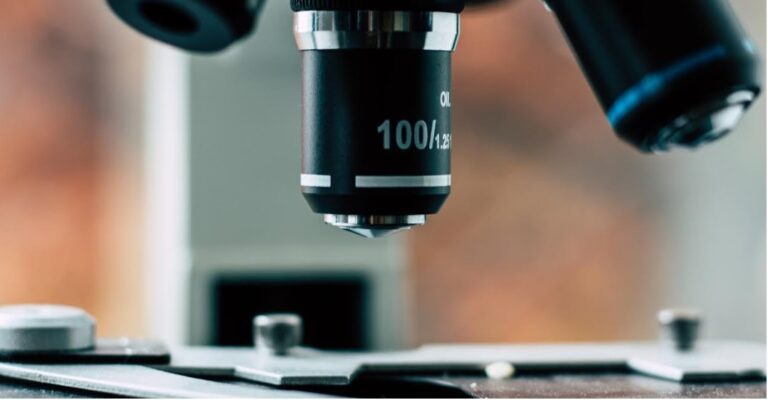
Artificial intelligence applications in microscopy are transforming how doctors and researchers analyze samples. AI is helping biology professionals overcome common hurdles like lengthy sample analysis, diagnosis delays, poor imaging quality and more.
Emerging AI-powered innovations could even make next-gen microscopy more accessible to doctors worldwide. Here’s how AI is transforming and improving microscopy and sample analysis.
Augmented Reality Microscopy
“What if doctors could look at a sample on their microscope and immediately view analysis data on the slide? That’s the goal of a cutting-edge collaboration between Google and the U.S.”
Department of Defense. The project integrates deep learning AI into a next-gen microscope for real-time sample analysis displayed in augmented reality.
Google designed this technology for rapid cancer diagnosis. The Augmented Reality Microscope, or ARM, analyzes cell samples in real time directly on the microscope. Then, it displays the analysis results as an AR overlay on the images. The doctor or researcher can view the sample and analyze data without transferring the images to a computer or mailing the sample for analysis. As a result, they can get a diagnosis within minutes rather than days.
The ARM project is still in its early stages since launching in 2020. The technology remains too expensive for widespread adoption. However, the plan is for ARMs to become more accessible down the road. In addition to decreasing in price, this technology could also be adapted to analyze different types of samples or diagnose more conditions.
What’s particularly exciting about this application of AI in microscopy is Google’s plans for the technology. Right now, the ARM prototypes are unique, specialized devices. In the future, Google aims to condense this technology into an attachment any doctor or researcher could add to their conventional optical microscope. With a single accessory, someone could transform it into a next-gen AI microscopy tool.
AI-Powered Computational Microscopy
Microscopes are incredible devices, but like any technology, they have limitations. A doctor may need to use a combination to get comprehensive results. For instance, a standard optical microscope might be fine for basic visual sample inspection, but a researcher might need a fluorescence microscope to gather more detailed data on different objects or molecules in their sample.
What if doctors and researchers could do everything from one microscope or do more with low-cost equipment? Many smaller labs and university researchers can’t afford to invest in expensive, specialized microscopes. AI can help them expand their capabilities so they can collect more data from a single microscope and rely less on costly equipment.
“Computational microscopy uses AI to enhance image quality without changing imaging hardware.”
Researchers can increase resolution, depth of field, contrast and more purely through AI software. As a result, they can capture more accurate, useful images with low-cost hardware.
Unlike some other emerging applications for AI in microscopy, computational imaging is already becoming accessible to the masses. It’s widely found in today’s consumer smartphones. For example, Apple uses AI software to improve and automate image quality and processing.
Researchers can do the same thing with microscopes. Computational microscopy could make cutting-edge imaging and sample analysis more accessible to doctors and researchers worldwide, particularly those without the funds for more advanced equipment.
Rapid, Automated Sample Analysis
Sample analysis is one of the top applications for AI in microscopy. These images can be complex and time-consuming to analyze, posing a major hurdle for small teams or situations requiring rapid results. For instance, analyzing samples for medical applications can significantly extend the time it takes to get a patient’s diagnosis, delaying the start of potentially life-saving treatment.
AI can resolve these challenges. AI and machine learning are fantastic tools for analyzing data. It’s repetitive and based on identifying consistent patterns and indicators, making it perfect for automation.
“Doctors and researchers can use AI to analyze microscopy images in a fraction of the time manual analysis would take and identify more trends and insights along the way. ”
For example, doctors use AI data analytics to speed up medicine discovery and development. They can have AI autonomously monitor changes in a sample over time or use computer vision to analyze images captured on a microscope. AI condenses tasks that would normally extend the research and development process by weeks or months. This allows researchers to develop a medicine far more quickly and with fewer resources and team members.
AI and the Future of Microscopy
Artificial intelligence is revolutionizing how researchers, scientists and doctors use their microscopes. With the help of AI, they can increase their microscopes’ capabilities, rapidly analyze samples, diagnose patients in minutes and streamline medicine development.
The applications for AI in microscopy could also one day make cutting-edge sample analysis accessible to the masses with low-cost, high-tech tools like AI microscope attachments. These scientific advances can lead to improved medical research and health care outcomes.